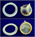
Aortic valve
The aortic valve is a valve in the heart of humans and most other animals, located between the left ventricle and the aorta. It is one of the four valves of the heart and one of the two semilunar valves, the other being the pulmonary valve. The aortic valve normally has three cusps or leaflets, although in 1–2% of the population it is found to congenitally have two leaflets.[1] The aortic valve is the last structure in the heart the blood travels through before stopping the flow through the systemic circulation.[1]
Aortic valve
Structure[edit]
The aortic valve normally has three cusps however there is some discrepancy in their naming.[2] They may be called the left coronary, right coronary and non-coronary cusp.[2] Some sources also advocate they be named as a left, right and posterior cusp.[3][4] Anatomists have traditionally named them the left posterior (origin of left coronary), anterior (origin of the right coronary) and right posterior.[2]
The three cusps, when the valve is closed, contain a sinus called an aortic sinus or sinus of Valsalva. In two of these cusps, the origin of the coronary arteries are found. The width of the sinuses in cross-section is wider than the left ventricular outflow tract as well as wider than the ascending aorta. The junction of the sinuses with the aorta is called the sinotubular junction. The aortic valve is located posterior to the pulmonary valve and the commissure where the anterior two cusps join together points toward the pulmonary valve. It is these two sinuses that contain the origin of the coronary arteries. In the congenital disease known as transposition of the great arteries, these two valves are reversed (the anterior valve is the aortic valve) and the origin of the coronaries still follows this "rule" that the origins are in the sinuses facing the pulmonary valve.
The term "semilunar" refers to an approximate half-moon shape of the valve leaflets.[5]


