
Cerebral cortex
The cerebral cortex, also known as the cerebral mantle,[1] is the outer layer of neural tissue of the cerebrum of the brain in humans and other mammals. It is the largest site of neural integration in the central nervous system,[2] and plays a key role in attention, perception, awareness, thought, memory, language, and consciousness. The cerebral cortex is the part of the brain responsible for cognition.
For the scientific journal, see Cerebral Cortex (journal). For the cerebellar cortex, see Cerebellum § Gross anatomy.Cerebral cortex
$_$_$DEEZ_NUTS#0__titleDEEZ_NUTS$_$_$
$_$_$DEEZ_NUTS#0__subtitleDEEZ_NUTS$_$_$
The six-layered neocortex makes up approximately 90% of the cortex, with the allocortex making up the remainder.[3] The cortex is divided into left and right parts by the longitudinal fissure, which separates the two cerebral hemispheres that are joined beneath the cortex by the corpus callosum. In most mammals, apart from small mammals that have small brains, the cerebral cortex is folded, providing a greater surface area in the confined volume of the cranium. Apart from minimising brain and cranial volume, cortical folding is crucial for the brain circuitry and its functional organisation.[4] In mammals with small brains, there is no folding and the cortex is smooth.[5][6]
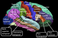
A fold or ridge in the cortex is termed a gyrus (plural gyri) and a groove is termed a sulcus (plural sulci). These surface convolutions appear during fetal development and continue to mature after birth through the process of gyrification. In the human brain, the majority of the cerebral cortex is not visible from the outside, but buried in the sulci.[7] The major sulci and gyri mark the divisions of the cerebrum into the lobes of the brain. The four major lobes are the frontal, parietal, occipital and temporal lobes. Other lobes are the limbic lobe, and the insular cortex often referred to as the insular lobe.
There are between 14 and 16 billion neurons in the human cerebral cortex.[2] These are organised into horizontal cortical layers, and radially into cortical columns and minicolumns. Cortical areas have specific functions such as movement in the motor cortex, and sight in the visual cortex. The motor cortex is primarily located in the precentral gyrus, and the visual cortex is located in the occipital lobe.
$_$_$DEEZ_NUTS#3__titleDEEZ_NUTS$_$_$
$_$_$DEEZ_NUTS#3__descriptionDEEZ_NUTS$_$_$
$_$_$DEEZ_NUTS#2__titleDEEZ_NUTS$_$_$
$_$_$DEEZ_NUTS#2__descriptionDEEZ_NUTS$_$_$
History[edit]
In 1909, Korbinian Brodmann distinguished different areas of the neocortex based on cytoarchitectural difference and divided the cerebral cortex into 52 regions.[80]
Rafael Lorente de Nó, a student of Santiago Ramon y Cajal identified more than 40 different types of cortical neurons based on the distribution of their dendrites and axons.[80]
Lateral surface of the human cerebral cortex
Medial surface of the human cerebral cortex

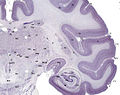
Tissue slice from the brain of an adult macaque monkey. The cerebral cortex is depicted in dark violet.
$_$_$DEEZ_NUTS#4__titleDEEZ_NUTS$_$_$
$_$_$DEEZ_NUTS#4__subtextDEEZ_NUTS$_$_$
$_$_$DEEZ_NUTS#1__titleDEEZ_NUTS$_$_$
$_$_$DEEZ_NUTS#1__subtextDEEZ_NUTS$_$_$
$_$_$DEEZ_NUTS#1__answer--0DEEZ_NUTS$_$_$
$_$_$DEEZ_NUTS#1__answer--1DEEZ_NUTS$_$_$
$_$_$DEEZ_NUTS#1__answer--2DEEZ_NUTS$_$_$
$_$_$DEEZ_NUTS#1__answer--3DEEZ_NUTS$_$_$
$_$_$DEEZ_NUTS#1__answer--4DEEZ_NUTS$_$_$
$_$_$DEEZ_NUTS#1__answer--5DEEZ_NUTS$_$_$
$_$_$DEEZ_NUTS#1__answer--6DEEZ_NUTS$_$_$