Iris (anatomy)
The iris (pl.: irides or irises) is a thin, annular structure in the eye in most mammals and birds, responsible for controlling the diameter and size of the pupil, and thus the amount of light reaching the retina. In optical terms, the pupil is the eye's aperture, while the iris is the diaphragm. Eye color is defined by the iris.
This article is about the part of the eye. For other uses, see Iris (disambiguation).Function[edit]
The iris controls the size of the pupil by means of contracting the iris sphincter muscle and/or the iris dilator muscle. The size of the pupils is dependent on many factors (including light, emotional state, cognitive load, arousal, stimulation), and can range from less than 2 mm in diameter, to as large as 9 mm in diameter. However, there is considerable variation in maximal pupil diameter by individual humans, and decreases with age.[6][7] The irises also contract the pupils when accommodation is initiated, to increase the depth of field.
Very few humans possess the ability to exert direct voluntary control over their iris muscles, which grants them the ability to dilate and constrict their pupils on command.[8] However, there is no clear purpose or advantage to this.
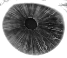
Iris, front view
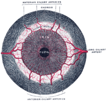
Fluorescein angiograpy of the iris reveals a radial layout of blood vessels.