Katana VentraIP

Karyotype

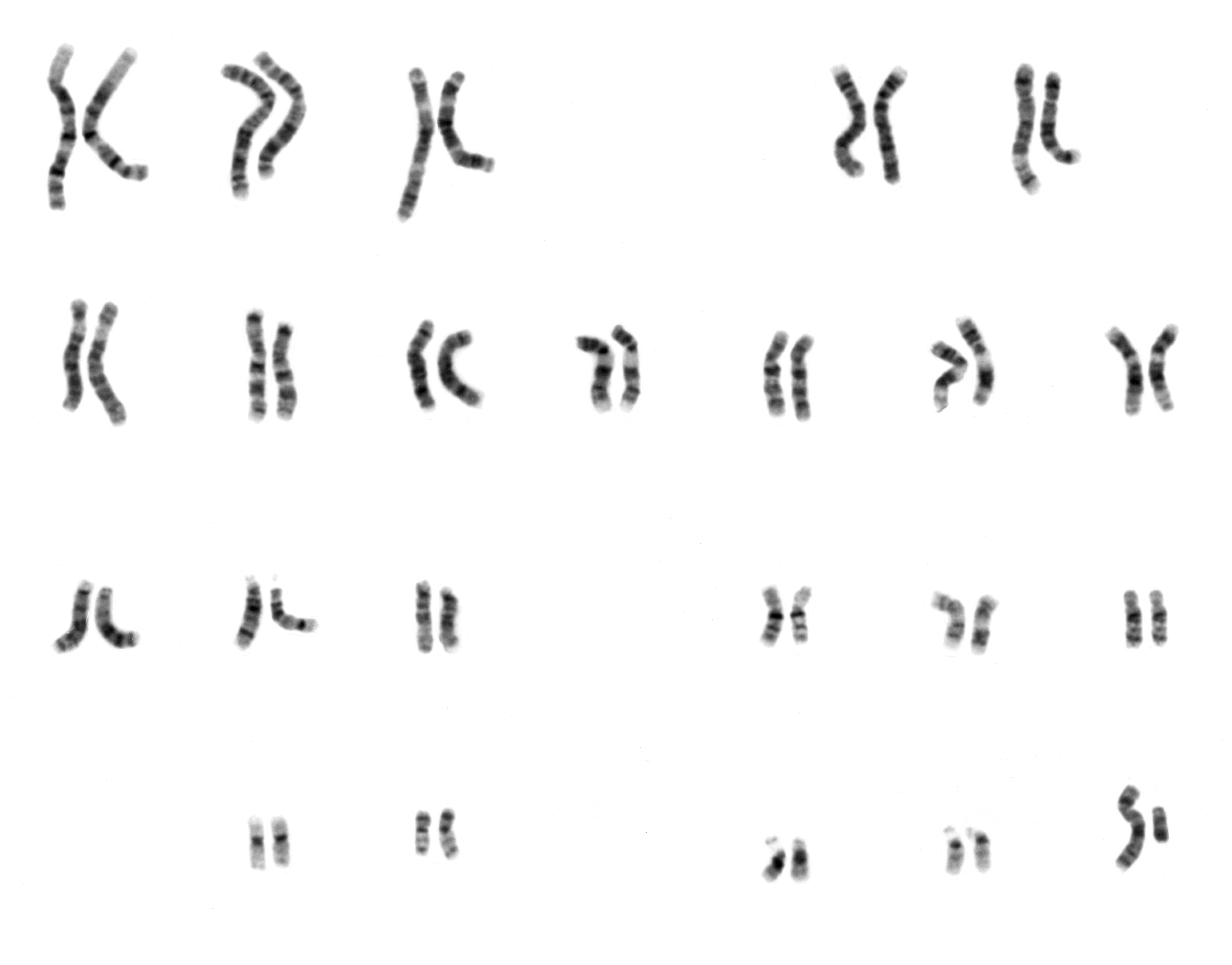
A karyotype is the general appearance of the complete set of chromosomes in the cells of a species or in an individual organism, mainly including their sizes, numbers, and shapes.[1][2] Karyotyping is the process by which a karyotype is discerned by determining the chromosome complement of an individual, including the number of chromosomes and any abnormalities.

$_$_$DEEZ_NUTS#1__subtextDEEZ_NUTS$_$_$

$_$_$DEEZ_NUTS#1__answer--0DEEZ_NUTS$_$_$

Katana VentraIP

$_$_$DEEZ_NUTS#0__titleDEEZ_NUTS$_$_$

$_$_$DEEZ_NUTS#0__subtitleDEEZ_NUTS$_$_$

"Idiogram" redirects here. Not to be confused with ideogram.

$_$_$DEEZ_NUTS#2__descriptionDEEZ_NUTS$_$_$

$_$_$DEEZ_NUTS#3__descriptionDEEZ_NUTS$_$_$

$_$_$DEEZ_NUTS#1__titleDEEZ_NUTS$_$_$

A karyogram or idiogram is a graphical depiction of a karyotype, wherein chromosomes are generally organized in pairs, ordered by size and position of centromere for chromosomes of the same size. Karyotyping generally combines light microscopy and photography in the metaphase of the cell cycle, and results in a photomicrographic (or simply micrographic) karyogram. In contrast, a schematic karyogram is a designed graphic representation of a karyotype. In schematic karyograms, just one of the sister chromatids of each chromosome is generally shown for brevity, and in reality they are generally so close together that they look as one on photomicrographs as well unless the resolution is high enough to distinguish them. The study of whole sets of chromosomes is sometimes known as karyology.


Karyotypes describe the chromosome count of an organism and what these chromosomes look like under a light microscope. Attention is paid to their length, the position of the centromeres, banding pattern, any differences between the sex chromosomes, and any other physical characteristics.[3] The preparation and study of karyotypes is part of cytogenetics.


The basic number of chromosomes in the somatic cells of an individual or a species is called the somatic number and is designated 2n. In the germ-line (the sex cells) the chromosome number is n (humans: n = 23).[4][5]p28 Thus, in humans 2n = 46.


So, in normal diploid organisms, autosomal chromosomes are present in two copies. There may, or may not, be sex chromosomes. Polyploid cells have multiple copies of chromosomes and haploid cells have single copies.


Karyotypes can be used for many purposes; such as to study chromosomal aberrations, cellular function, taxonomic relationships, medicine and to gather information about past evolutionary events (karyosystematics).[6]

Depiction of karyotypes[edit]

Types of banding[edit]

Cytogenetics employs several techniques to visualize different aspects of chromosomes:[9]

$_$_$DEEZ_NUTS#4__titleDEEZ_NUTS$_$_$

$_$_$DEEZ_NUTS#4__descriptionDEEZ_NUTS$_$_$

$_$_$DEEZ_NUTS#4__heading--0DEEZ_NUTS$_$_$

$_$_$DEEZ_NUTS#4__description--0DEEZ_NUTS$_$_$

$_$_$DEEZ_NUTS#4__heading--1DEEZ_NUTS$_$_$

$_$_$DEEZ_NUTS#4__description--1DEEZ_NUTS$_$_$

$_$_$DEEZ_NUTS#4__heading--2DEEZ_NUTS$_$_$

$_$_$DEEZ_NUTS#4__description--2DEEZ_NUTS$_$_$

$_$_$DEEZ_NUTS#4__heading--3DEEZ_NUTS$_$_$

$_$_$DEEZ_NUTS#4__description--3DEEZ_NUTS$_$_$

results from a single X chromosome (45,X or 45,X0).

Turner syndrome

the most common male chromosomal disease, otherwise known as 47,XXY, is caused by an extra X chromosome.

Klinefelter syndrome

is caused by trisomy (three copies) of chromosome 18.

Edwards syndrome

$_$_$DEEZ_NUTS#2__titleDEEZ_NUTS$_$_$

22 autosomal chromosome pairs (chromosomes 1 to 22). Homologous means that they have the same genes in the same loci, and autosomal means that they are not sex chromomes.

homologous

Two (in green rectangle at bottom right in the schematic karyogram, with adjacent silhouettes of typical representative phenotypes): The most common karyotypes for females contain two X chromosomes and are denoted 46,XX; males usually have both an X and a Y chromosome denoted 46,XY. However, approximately 0.018% percent of humans are intersex, sometimes due to variations in sex chromosomes.[16]

sex chromosome

The (shown at bottom left in the schematic karyogram, to scale compared to the nuclear DNA in terms of base pairs), although this is not included in micrographic karyograms in clinical practice. Its genome is relatively tiny compared to the rest.

human mitochondrial genome

Chromosome elimination. In some species, as in many , entire chromosomes are eliminated during development.[22]

sciarid flies

Chromatin diminution (founding father: ). In this process, found in some copepods and roundworms such as Ascaris suum, portions of the chromosomes are cast away in particular cells. This process is a carefully organised genome rearrangement where new telomeres are constructed and certain heterochromatin regions are lost.[23][24] In A. suum, all the somatic cell precursors undergo chromatin diminution.[25]

Theodor Boveri

. The inactivation of one X chromosome takes place during the early development of mammals (see Barr body and dosage compensation). In placental mammals, the inactivation is random as between the two Xs; thus the mammalian female is a mosaic in respect of her X chromosomes. In marsupials it is always the paternal X which is inactivated. In human females some 15% of somatic cells escape inactivation,[26] and the number of genes affected on the inactivated X chromosome varies between cells: in fibroblast cells up about 25% of genes on the Barr body escape inactivation.[27]

X-inactivation

$_$_$DEEZ_NUTS#5__titleDEEZ_NUTS$_$_$

$_$_$DEEZ_NUTS#5__subtextDEEZ_NUTS$_$_$

$_$_$DEEZ_NUTS#5__quote--0DEEZ_NUTS$_$_$

$_$_$DEEZ_NUTS#5__name--0DEEZ_NUTS$_$_$

$_$_$DEEZ_NUTS#5__company_or_position--0DEEZ_NUTS$_$_$

$_$_$DEEZ_NUTS#5__quote--1DEEZ_NUTS$_$_$

$_$_$DEEZ_NUTS#5__name--1DEEZ_NUTS$_$_$

$_$_$DEEZ_NUTS#5__company_or_position--1DEEZ_NUTS$_$_$

$_$_$DEEZ_NUTS#5__quote--2DEEZ_NUTS$_$_$

$_$_$DEEZ_NUTS#5__name--2DEEZ_NUTS$_$_$

$_$_$DEEZ_NUTS#5__company_or_position--2DEEZ_NUTS$_$_$

a common chromosomal disease, is caused by trisomy of chromosome 21.

Down syndrome

is caused by trisomy of chromosome 13.

Patau syndrome

believed to be the 4th most common trisomy, has many long lived affected individuals but only in a form other than a full trisomy, such as trisomy 9p syndrome or mosaic trisomy 9. They often function quite well, but tend to have trouble with speech.

Trisomy 9

Also documented are trisomy 8 and trisomy 16, although they generally do not survive to birth.

 – Symbols and abbreviations used in cytogenetics

Cytogenetic notation

 – Laboratory process

Genome screen

Media related to Karyotypes at Wikimedia Commons

an online activity from the University of Utah's Genetic Science Learning Center.

Making a karyotype

from the University of Arizona's Biology Project.

Karyotyping activity with case histories

from Biology Corner, a resource site for biology and science teachers.

Printable karyotype project

Chromosome Staining and Banding Techniques

Archived 12 June 2019 at the Wayback Machine

Bjorn Biosystems for Karyotyping and FISH