Katana VentraIP

Renal vein

The renal veins in the renal circulation, are large-calibre[1] veins that drain blood filtered by the kidneys into the inferior vena cava. There is one renal vein draining each kidney. Each renal vein is formed by the convergence of the interlobar veins of one kidney.[2]

Because the inferior vena cava is on the right half of the body, the left renal vein is longer than the right one.

the anterior branch which receives blood from the anterior portion of the kidney and,

the posterior branch which receives blood from the posterior portion.

Clinical significance[edit]

Diseases associated with the renal vein include renal vein thrombosis (RVT) and nutcracker syndrome (renal vein entrapment syndrome).

3D-rendered computed tomography, showing one renal vein (in red color) for each kidney

3D-rendered computed tomography, showing one renal vein (in red color) for each kidney

Frontal section through the kidney

Frontal section through the kidney

Diagram showing completion of development of the parietal veins.

Diagram showing completion of development of the parietal veins.

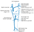
The venæ cavæ and azygos veins, with their tributaries.

The venæ cavæ and azygos veins, with their tributaries.

Renal vein

Renal vein

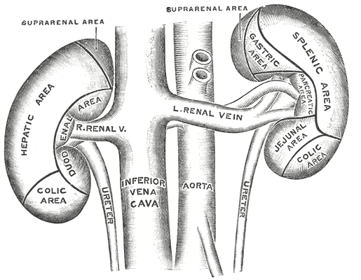
Human kidneys viewed from behind with spine removed.

Human kidneys viewed from behind with spine removed.

Kidney

Kidney

Renal vein

Renal vein

Renal vein

Renal vein

Renal vein

Renal vein

Renal physiology

Nutcracker syndrome

Renal artery

at Human Anatomy Online, SUNY Downstate Medical Center - "Retroperitoneal structures on the posterior abdominal wall."

Anatomy figure: 40:06-05