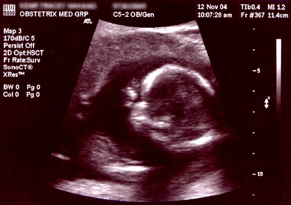
Obstetric ultrasonography
Obstetric ultrasonography, or prenatal ultrasound, is the use of medical ultrasonography in pregnancy, in which sound waves are used to create real-time visual images of the developing embryo or fetus in the uterus (womb). The procedure is a standard part of prenatal care in many countries, as it can provide a variety of information about the health of the mother, the timing and progress of the pregnancy, and the health and development of the embryo or fetus.
The International Society of Ultrasound in Obstetrics and Gynecology (ISUOG) recommends that pregnant women have routine obstetric ultrasounds between 18 weeks' and 22 weeks' gestational age (the anatomy scan) in order to confirm pregnancy dating, to measure the fetus so that growth abnormalities can be recognized quickly later in pregnancy, and to assess for congenital malformations and multiple pregnancies (twins, etc).[1] Additionally, the ISUOG recommends that pregnant patients who desire genetic testing have obstetric ultrasounds between 11 weeks' and 13 weeks 6 days' gestational age in countries with resources to perform them (the nuchal scan). Performing an ultrasound at this early stage of pregnancy can more accurately confirm the timing of the pregnancy, and can also assess for multiple fetuses and major congenital abnormalities at an earlier stage.[2] Research shows that routine obstetric ultrasound before 24 weeks' gestational age can significantly reduce the risk of failing to recognize multiple gestations and can improve pregnancy dating to reduce the risk of labor induction for post-dates pregnancy. There is no difference, however, in perinatal death or poor outcomes for infants.[3]
Below are useful terms on ultrasound:[4]
In normal state, each body tissue type, such as liver, spleen or kidney, has a unique echogenicity. Fortunately, gestational sac, yolk sac and embryo are surrounded by hyperechoic (brighter) body tissues.
Medical uses[edit]
Early pregnancy[edit]
A gestational sac can be reliably seen on transvaginal ultrasound by 5 weeks' gestational age (approximately 3 weeks after ovulation). The embryo should be seen by the time the gestational sac measures 25 mm, about five and a half weeks.[10] The heartbeat is usually seen on transvaginal ultrasound by the time the embryo measures 5 mm, but may not be visible until the embryo reaches 19 mm, around 7 weeks' gestational age.[5][11][12] Coincidentally, most miscarriages also happen by 7 weeks' gestation. The rate of miscarriage, especially threatened miscarriage, drops significantly after normal heartbeat is detected, and after 13 weeks.[13]
Society and culture[edit]
The increasingly widespread use of ultrasound technology in monitoring pregnancy has had a great impact on the way in which women and societies at large conceptualise and experience pregnancy and childbirth.[37] The pervasive spread of obstetric ultrasound technology around the world and the conflation of its use with creating a 'safe' pregnancy as well as the ability to see and determine features like the sex of the fetus affect the way in which pregnancy is experienced and conceptualised.[37] This "technocratic takeover"[37] of pregnancy is not limited to western or developed nations but also affects conceptualisations and experiences in developing nations and is an example of the increasing medicalisation of pregnancy, a phenomenon that has social as well as technological ramifications.[37] Ethnographic research concerned with the use of ultrasound technology in monitoring pregnancy can show us how it has changed the embodied experience of expecting mothers around the globe.[37]
Recent studies have stressed the importance of framing "reproductive health matters cross-culturally", particularly when understanding the "new phenomenon" of "the proliferation of ultrasound imaging" in developing countries.[38] In 2004, Tine Gammeltoft interviewed 400 women in Hanoi's Obstetrics and Gynecology Hospital; each "had an average of 6.6 scans during her pregnancy", much higher than five years prior when "a pregnant woman might or might not have had a single scan during her pregnancy" in Vietnam.[38] Gammeltoft explains that "many Asian countries" see "the foetus as an ambiguous being" unlike in Western medicine where it is common to think of the foetus as "materially stable".[38] Therefore, although women, particularly in Asian countries, "express intense uncertainties regarding the safety and credibility of this technology", it is overused for its "immediate reassurance".[38]


