
Gall
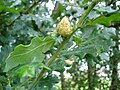
Galls (from the Latin galla, 'oak-apple') or cecidia (from the Greek kēkidion, anything gushing out) are a kind of swelling growth on the external tissues of plants. Plant galls are abnormal outgrowths[1] of plant tissues, similar to benign tumors or warts in animals. They can be caused by various parasites, from viruses, fungi and bacteria, to other plants, insects and mites. Plant galls are often highly organized structures so that the cause of the gall can often be determined without the actual agent being identified. This applies particularly to insect and mite plant galls. The study of plant galls is known as cecidology.
This article is about the abnormal growths in plants. For other uses, see Gall (disambiguation).Anatomy[edit]
Shape and size[edit]
Galls develop on various plant organs, providing nutrition and shelter to inducing insects. Galls display vast variation in morphology, size, and wall composition. The size of insect galls can range significantly, from approximately two inches in diameter to less than one-sixteenth of an inch. Some galls are so small that they are merely slightly thickened patches on leaves.[2] Their shape can range from spherical to bursiform, bullet-shaped, flower-shaped, cylindrical, or diamond-like. Factors influencing gall morphology include plant species, tissue type, gall-inducing agent, and environmental conditions.[3][4][5][6][7] They typically exhibit symmetrical forms, although their end shapes vary due to differences in the physical actions and chemical stimuli of different insects. Around 90% of galls occur on the leaves of dicotyledons.[8] Galls can develop on various parts of the host plant, such as roots, leaf bases, branches, or leaflets. Internally, galls also exhibit diverse structures. Some are simple, comprising only outgrown and curved leaf tissues, while others feature complex, hierarchical arrangements with multiple chambers containing different types of tissues, including collenchyma, parenchyma, physalides-parenchyma, and a nutritive cellular layer.[9][10][11]
Structure[edit]
In a general gall wasp gall, the outermost layer is the epidermis followed by outer cortex and then inner cortex. In some galls these two cortex layers are separated by a lignified layer. The innermost part of a gall is the larval chamber. The nutritive layer situated between the larval chamber and the inner cortex. There is a nutritional gradient (high to low) from inside to outside of the gall while defense gradient to the opposite direction.[12][13]
Morphogenesis[edit]
Gall morphogenesis involves the regulation of the organ on which the gall occurs while maintaining differentiation freedom. Gall development begins from a single or group of metaplasied cells and progresses through promoter-mediated cell expansion, cell multiplication, programmed differentiation, and control of symmetry.[8]
Plant response involves the establishment of metaplasied cells and localized metabolic changes to repair the wound and neutralize stress. Osmotic stress leads to the development of metaplasied cells, characterized by increased quantities of osmotically active material. The rejection response by the plant triggers the synthesis of defense compounds and enzymes.[14][15]
Genetics[edit]
Galls are unique growths on plants, and how the plant's genetic instructions could produce these structures in response to external factors is still a fresh field of science. Genetic mechanisms of gall formation is a unique interplay between the parasite and the host plant in shaping the developmental trajectory of the gall organ.
The 'zigzag' model introduced by Jones & Dangl (2006)[26] demonstrates the molecular interactions underlying gall induction. This model, refined over time and subject to ongoing enhancements, illustrates the intricate dynamics between antagonistic molecular players. Pattern-triggered immunity (PTI), constitutes the initial defense layer of plant cells, activated upon detection of "danger signals." These signals, termed damage-associated-molecular-patterns (DAMPs) if originating from the plant or microbe/pathogen-associated-molecular-patterns (MAMPs, PAMPs, or HAMPs)[27] if from the parasite, engage pattern-recognition receptors (PRRs) triggering signaling cascades. PRRs, classified as receptor-like kinases (RLKs), mediate intercellular communication by bridging external stimuli with intracellular defense mechanisms. Antagonists, employing effector-triggered susceptibility (ETS) manipulate host-cell functions through effector molecules encoded by effector genes, aiming primarily at suppressing plant defenses. Notably, some effectors exploit plant traits, known as "plant susceptibility traits," diverting the plant's resources in favor of the parasite. Effectoromics, involving high-throughput expression screens, aids in identifying effector candidates crucial for colonization. Conversely, Effector-Triggered Immunity (ETI) responsible for plant's counterattack, leveraging effectors as "danger signals" to render the parasite avirulent. During ETI, nucleotide-binding domain leucine-rich repeat (NLR)-containing receptors detect perturbations induced by effectors, leading to downstream signaling events that promote defense responses. However, parasites can counteract ETI by modifying ETS, undermining the efficacy of resistance genes deployed in agriculture. The evolutionary arms race between plants and parasites, underscored by the expansion of gene families involved in biotic interactions, shapes their genomic landscape, influencing their adaptive strategies and diversification.[28][29]
Crown galls formed under the influence of the bacterium Agrobacterium tumefaciens exhibit several distinctive characteristics when compared to other types of galls. This bacterium transfers genetic material known as T-DNA into the plant cells, where it becomes integrated into the chromosomes. The T-DNA contains genes that encode for production of auxin, cytokinin and opines. As a result, the infected plant cells undergo rapid multiplication, essentially transforming into "bacterial factories" that produce more bacterial bodies.[2]
Certain bacteria, like Rhodococcus fascians, induce the formation of leafy galls on plants, affecting their growth. These galls act as permanent sinks, diverting nutrients away from other parts of the plant and causing growth suppression elsewhere. The bacteria possess virulence genes that control their ability to colonize plants and produce cytokinins, which influence plant growth. While parasitic gall-inducers are typically harmful to plants, researchers are exploring ways to harness their growth-promoting abilities for agricultural benefit. Some derivatives of R. fascians are being investigated for their potential to promote balanced plant growth, and scientists are also studying plant interactions with these bacteria to discover traits that could enhance crop yields.
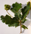
Most of the transcriptomic studies on plant galls used entire gall samples resulting both gall and non-gall cells leading to thousands of gene expressions during gall development.[30][31] Recent studies on gall induced by gall wasps (Hymenoptera: Cynipidae)[32] Dryocosmus quercuspalustris on northern red oak (Quercus rubra L.) leaves demonstrate the complexity of genetic mechanisms underlying galls by quantifying the tissue-specific gene expression.[33] There are substantial differences in gene expression between inner and outer gall tissues compared to adjacent leaf tissues. Notably, approximately 28% of oak genes display differential expression in the gall compared to leaves, indicating significant transcriptional changes associated with gall development.[33] According to the transcriptome analysis, while the outer gall transcriptome resembles that of twigs, leaf buds, and reproductive structures, the inner gall transcriptome is distinct from normal oak tissues, underscoring the complexity of gall formation.[12] Furthermore, there is an upregulation of genes related to sugar and amino acid metabolism in both outer and inner gall tissues, suggesting a role in transporting plant metabolites to support the nutritional needs of the developing gall wasp larva. The defense-related genes are found to be suppressed in inner gall tissues as a strategy to accommodate the feeding activity of the parasite.[34]
Artichoke gall cut open to reveal wasp larva
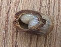


Cherry oak gall cut open to reveal wasp larva

Cherry oak gall wasp adult


Uses[edit]
Galls are rich in resins and tannic acid and have been used widely in the manufacturing of permanent inks (such as iron gall ink) and astringent ointments, in dyeing, and in leather tanning. The Talmud[43] records using gallnuts as part of the tanning process as well as a dye-base for ink.
Medieval Arabic literature records many uses for the gall, called ˁafṣ in Arabic. The Aleppo gall, found on oak trees in northern Syria, was among the most important exports from Syria during this period, with one merchant recording a shipment of galls from Suwaydiyya near Antioch fetching the high price of 4½ dinars per 100 pounds. The primary use of the galls was as a mordant for black dyes; they were also used to make a high-quality ink. The gall was also used as a medication to treat fever and intestinal ailments.[44]