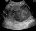
Uterine fibroid
Uterine fibroids, also known as uterine leiomyomas or fibroids, are benign smooth muscle tumors of the uterus, part of the female reproductive system.[1] Some women with fibroids have no symptoms while others may have painful or heavy periods.[1] If large enough, they may push on the bladder, causing a frequent need to urinate.[1] They may also cause pain during penetrative sex or lower back pain.[1][3] A woman can have one uterine fibroid or many.[1] It is uncommon but possible that fibroids may make it difficult to become pregnant.[1]
Not to be confused with Leiomyosarcoma.Uterine fibroids
Uterine leiomyoma, uterine myoma, myoma, fibromyoma, fibroleiomyoma
Middle and later reproductive years[1]
Unknown[1]
Medications, surgery, uterine artery embolization[1]
Ibuprofen, paracetamol (acetaminophen), iron supplements, gonadotropin releasing hormone agonist[1]
~50% of women by age 50[1]
The exact cause of uterine fibroids is unclear.[1] However, fibroids run in families and appear to be partly determined by hormone levels.[1] Risk factors include obesity and eating red meat.[1] Diagnosis can be performed by pelvic examination or medical imaging.[1]
Treatment is typically not needed if there are no symptoms.[1] NSAIDs, such as ibuprofen, may help with pain and bleeding while paracetamol (acetaminophen) may help with pain.[1][4] Iron supplements may be needed in those with heavy periods.[1] Medications of the gonadotropin-releasing hormone agonist class may decrease the size of the fibroids but are expensive and associated with side effects.[1] If greater symptoms are present, surgery to remove the fibroid or uterus may help.[1] Uterine artery embolization may also help.[1] Cancerous versions of fibroids are very rare and are known as leiomyosarcomas.[1] They do not appear to develop from benign fibroids.[1]
About 20% to 80% of women develop fibroids by the age of 50.[1] In 2013, it was estimated that 171 million women were affected worldwide.[5] They are typically found during the middle and later reproductive years.[1] After menopause, they usually decrease in size.[1] In the United States, uterine fibroids are a common reason for surgical removal of the uterus.[6]
Signs and symptoms[edit]
Some women with uterine fibroids do not have symptoms. Abdominal pain, anemia and increased bleeding can indicate the presence of fibroids.[7] There may also be pain during intercourse (penetration), depending on the location of the fibroid. During pregnancy, they may also be the cause of miscarriage,[8] bleeding, premature labor, or interference with the position of the fetus.[9] A uterine fibroid can cause rectal pressure. The abdomen can grow larger mimicking the appearance of pregnancy.[1] Some large fibroids can extend out through the cervix and vagina.[7]
While fibroids are common, they are not a typical cause for infertility, accounting for about 3% of reasons why a woman may not be able to have a child.[10] The majority of women with uterine fibroids will have normal pregnancy outcomes.[11] In cases of intercurrent uterine fibroids in infertility, a fibroid is typically located in a submucosal position and it is thought that this location may interfere with the function of the lining and the ability of the embryo to implant.[10]
Related legislation[edit]
United States[edit]
The 2005 S.1289 bill was read twice and referred to the committee on Health, Labor, and Pensions but never passed for a Senate or House vote; the proposed Uterine Fibroid Research and Education Act of 2005 mentioned that $5 billion is spent annually on hysterectomy surgeries each year, which affect 22% of African Americans and 7% of Caucasian women. The bill also called for more funding for research and educational purposes. It also states that of the $28 billion issued to NIH, $5 million was allocated for uterine fibroids in 2004.[77]
Other animals[edit]
Uterine fibroids are rare in other mammals, although they have been observed in certain dogs and Baltic grey seals.[78]
Research[edit]
Selective progesterone receptor modulators, such as progenta, have been under investigation. Another selective progesterone receptor modulator asoprisnil is being tested with promising results as a possible use as a treatment for fibroids, intended to provide the advantages of progesterone antagonists without their adverse effects.[44] Low dietary intake of vitamin D is associated with the development of uterine fibroids.[12]


![Histopathology of uterine fibroids typically show smooth muscle in a whorled (fascicular) pattern.[39]](http://upload.wikimedia.org/wikipedia/commons/thumb/7/79/Histopathology_of_uterine_leiomyoma.jpg/120px-Histopathology_of_uterine_leiomyoma.jpg)

