
Vertebra
Each vertebra (pl.: vertebrae) is an irregular bone with a complex structure composed of bone and some hyaline cartilage, that make up the vertebral column or spine, of vertebrates. The proportions of the vertebrae differ according to their spinal segment and the particular species.
For other uses, see Vertebra (disambiguation).Vertebra
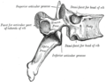
The basic configuration of a vertebra varies; the bone is the body, and the central part of the body
is the centrum. The upper and lower surfaces of the vertebra body give attachment to the intervertebral discs. The posterior part of a vertebra forms a vertebral arch, in eleven parts, consisting of two pedicles (pedicle of vertebral arch), two laminae, and seven processes. The laminae give attachment to the ligamenta flava (ligaments of the spine). There are vertebral notches formed from the shape of the pedicles, which form the intervertebral foramina when the vertebrae articulate. These foramina are the entry and exit conduits for the spinal nerves. The body of the vertebra and the vertebral arch form the vertebral foramen, the larger, central opening that accommodates the spinal canal, which encloses and protects the spinal cord.
Vertebrae articulate with each other to give strength and flexibility to the spinal column, and the shape at their back and front aspects determines the range of movement. Structurally, vertebrae are essentially alike across the vertebrate species, with the greatest difference seen between an aquatic animal and other vertebrate animals. As such, vertebrates take their name from the vertebrae that compose the vertebral column.

Vertebral joint

Functions of vertebrae include:
Clinical significance[edit]
There are a number of congenital vertebral anomalies, mostly involving variations in the shape or number of vertebrae, and many of which are unproblematic. Others though can cause compression of the spinal cord. Wedge-shaped vertebrae, called hemivertebrae can cause an angle to form in the spine which can result in the spinal curvature diseases of kyphosis, scoliosis and lordosis. Severe cases can cause spinal cord compression. Block vertebrae where some vertebrae have become fused can cause problems. Spina bifida can result from the incomplete formation of the vertebral arch.
Spondylolysis is a defect in the pars interarticularis of the vertebral arch. In most cases this occurs in the lowest of the lumbar vertebrae (L5), but may also occur in the other lumbar vertebrae, as well as in the thoracic vertebrae.
Spinal disc herniation, more commonly called a slipped disc, is the result of a tear in the outer ring (anulus fibrosus) of the intervertebral disc, which lets some of the soft gel-like material, the nucleus pulposus, bulge out in a hernia. This may be treated by a minimally-invasive endoscopic procedure called Tessys method.
A laminectomy is a surgical operation to remove the laminae in order to access the spinal canal.[21] The removal of just part of a lamina is called a laminotomy.
A pinched nerve caused by pressure from a disc, vertebra or scar tissue might be remedied by a foraminotomy to broaden the intervertebral foramina and relieve pressure. It can also be caused by a foramina stenosis, a narrowing of the nerve opening, as a result of arthritis.
Another condition is spondylolisthesis when one vertebra slips forward onto another. The reverse of this condition is retrolisthesis where one vertebra slips backward onto another.
The vertebral pedicle is often used as a radiographic marker and entry point in vertebroplasty, kyphoplasty, and spinal fusion procedures.
The arcuate foramen is a common anatomical variation more frequently seen in females. It is a bony bridge found on the first cervical vertebra, the atlas where it covers the groove for the vertebral artery.[22]
Degenerative disc disease is a condition usually associated with ageing in which one or more discs degenerate. This can often be a painfree condition but can also be very painful.
Vertebral arches of three thoracic vertebrae
Costovertebral joints seen from the front

Lower thoracic and upper lumbar vertebrae seen from the side

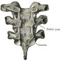
Cervical vertebrae seen from the back
