DNA vaccine
A DNA vaccine is a type of vaccine that transfects a specific antigen-coding DNA sequence into the cells of an organism as a mechanism to induce an immune response.[1][2]
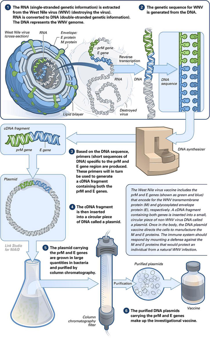
See also: messenger RNA (mRNA) vaccineDNA vaccines work by injecting genetically engineered plasmid containing the DNA sequence encoding the antigen(s) against which an immune response is sought, so the cells directly produce the antigen, thus causing a protective immunological response.[3] DNA vaccines have theoretical advantages over conventional vaccines, including the "ability to induce a wider range of types of immune response".[4] Several DNA vaccines have been tested for veterinary use.[3] In some cases, protection from disease in animals has been obtained, in others not.[3] Research is ongoing over the approach for viral, bacterial and parasitic diseases in humans, as well as for cancers.[4] In August 2021, Indian authorities gave emergency approval to ZyCoV-D. Developed by Cadila Healthcare, it is the first DNA vaccine approved for humans.[5]
Applications[edit]
As of 2021 no DNA vaccines have been approved for human use in the United States. Few experimental trials have evoked a response strong enough to protect against disease and the technique's usefulness remains to be proven in humans.
A veterinary DNA vaccine to protect horses from West Nile virus has been approved.[14] Another West Nile virus vaccine has been tested successfully on American robins.[15]
DNA immunization is also being investigated as a means of developing antivenom sera.[1] DNA immunization can be used as a technology platform for monoclonal antibody induction.[2]
Plasmid vectors[edit]
Vector design[edit]
DNA vaccines elicit the best immune response when high-expression vectors are used. These are plasmids that usually consist of a strong viral promoter to drive the in vivo transcription and translation of the gene (or complementary DNA) of interest.[18] Intron A may sometimes be included to improve mRNA stability and hence increase protein expression.[19] Plasmids also include a strong polyadenylation/transcriptional termination signal, such as bovine growth hormone or rabbit beta-globulin polyadenylation sequences.[6][7][20] Polycistronic vectors (with multiple genes of interest) are sometimes constructed to express more than one immunogen, or to express an immunogen and an immunostimulatory protein.[21]
Because the plasmid – carrying relatively small genetic code up to about 200 Kbp – is the "vehicle" from which the immunogen is expressed, optimising vector design for maximal protein expression is essential.[21] One way of enhancing protein expression is by optimising the codon usage of pathogenic mRNAs for eukaryotic cells. Pathogens often have different AT-contents than the target species, so altering the gene sequence of the immunogen to reflect the codons more commonly used in the target species may improve its expression.[22]
Another consideration is the choice of promoter. The SV40 promoter was conventionally used until research showed that vectors driven by the Rous Sarcoma Virus (RSV) promoter had much higher expression rates.[6] More recently, expression and immunogenicity have been further increased in model systems by the use of the cytomegalovirus (CMV) immediate early promoter, and a retroviral cis-acting transcriptional element.[23] Additional modifications to improve expression rates include the insertion of enhancer sequences, synthetic introns, adenovirus tripartite leader (TPL) sequences and modifications to the polyadenylation and transcriptional termination sequences.[6] An example of DNA vaccine plasmid is pVAC, which uses SV40 promoter.
Structural instability phenomena are of particular concern for plasmid manufacture, DNA vaccination and gene therapy.[24] Accessory regions pertaining to the plasmid backbone may engage in a wide range of structural instability phenomena. Well-known catalysts of genetic instability include direct, inverted and tandem repeats, which are conspicuous in many commercially available cloning and expression vectors. Therefore, the reduction or complete elimination of extraneous noncoding backbone sequences would pointedly reduce the propensity for such events to take place and consequently the overall plasmid's recombinogenic potential.[25]
Mechanism of plasmids[edit]
Once the plasmid inserts itself into the transfected cell nucleus, it codes for a peptide string of a foreign antigen. On its surface the cell displays the foreign antigen with both histocompatibility complex (MHC) classes I and class II molecules. The antigen-presenting cell then travels to the lymph nodes and presents the antigen peptide and costimulatory molecule signalling to T-cell, initiating the immune response.[26]
Vaccine insert design[edit]
Immunogens can be targeted to various cellular compartments to improve antibody or cytotoxic T-cell responses. Secreted or plasma membrane-bound antigens are more effective at inducing antibody responses than cytosolic antigens, while cytotoxic T-cell responses can be improved by targeting antigens for cytoplasmic degradation and subsequent entry into the major histocompatibility complex (MHC) class I pathway.[7] This is usually accomplished by the addition of N-terminal ubiquitin signals.[27][28][29]
The conformation of the protein can also affect antibody responses. "Ordered" structures (such as viral particles) are more effective than unordered structures.[30] Strings of minigenes (or MHC class I epitopes) from different pathogens raise cytotoxic T-cell responses to some pathogens, especially if a TH epitope is also included.[7]
Dosage[edit]
The delivery method determines the dose required to raise an effective immune response. Saline injections require variable amounts of DNA, from 10 μg to 1 mg, whereas gene gun deliveries require 100 to 1000 times less.[40] Generally, 0.2 μg – 20 μg are required, although quantities as low as 16 ng have been reported.[6] These quantities vary by species. Mice for example, require approximately 10 times less DNA than primates.[7] Saline injections require more DNA because the DNA is delivered to the extracellular spaces of the target tissue (normally muscle), where it has to overcome physical barriers (such as the basal lamina and large amounts of connective tissue) before it is taken up by the cells, while gene gun deliveries drive/force DNA directly into the cells, resulting in less "wastage".[6][7]
Mechanistic basis for DNA-raised immune responses[edit]
DNA uptake mechanism[edit]
When DNA uptake and subsequent expression was first demonstrated in vivo in muscle cells,[54] these cells were thought to be unique because of their extensive network of T-tubules. Using electron microscopy, it was proposed that DNA uptake was facilitated by caveolae (or, non-clathrin coated pits).[55] However, subsequent research revealed that other cells (such as keratinocytes, fibroblasts and epithelial Langerhans cells) could also internalize DNA.[46][56] The mechanism of DNA uptake is not known.
Two theories dominate – that in vivo uptake of DNA occurs non-specifically, in a method similar to phago- or pinocytosis,[21] or through specific receptors.[57] These might include a 30kDa surface receptor, or macrophage scavenger receptors. The 30kDa surface receptor binds specifically to 4500-bp DNA fragments (which are then internalised) and is found on professional APCs and T-cells. Macrophage scavenger receptors bind to a variety of macromolecules, including polyribonucleotides and are thus candidates for DNA uptake.[57][58] Receptor-mediated DNA uptake could be facilitated by the presence of polyguanylate sequences. Gene gun delivery systems, cationic liposome packaging, and other delivery methods bypass this entry method, but understanding it may be useful in reducing costs (e.g. by reducing the requirement for cytofectins), which could be important in animal husbandry.
Immune response modulation[edit]
Cytokine modulation[edit]
An effective vaccine must induce an appropriate immune response for a given pathogen. DNA vaccines can polarise T-cell help towards TH1 or TH2 profiles and generate CTL and/or antibody when required. This can be accomplished by modifications to the form of antigen expressed (i.e. intracellular vs. secreted), the method and route of delivery or the dose.[41][42][65][66][67] It can also be accomplished by the co-administration of plasmid DNA encoding immune regulatory molecules, i.e. cytokines, lymphokines or co-stimulatory molecules. These "genetic adjuvants" can be administered as a: