Mycobacterium
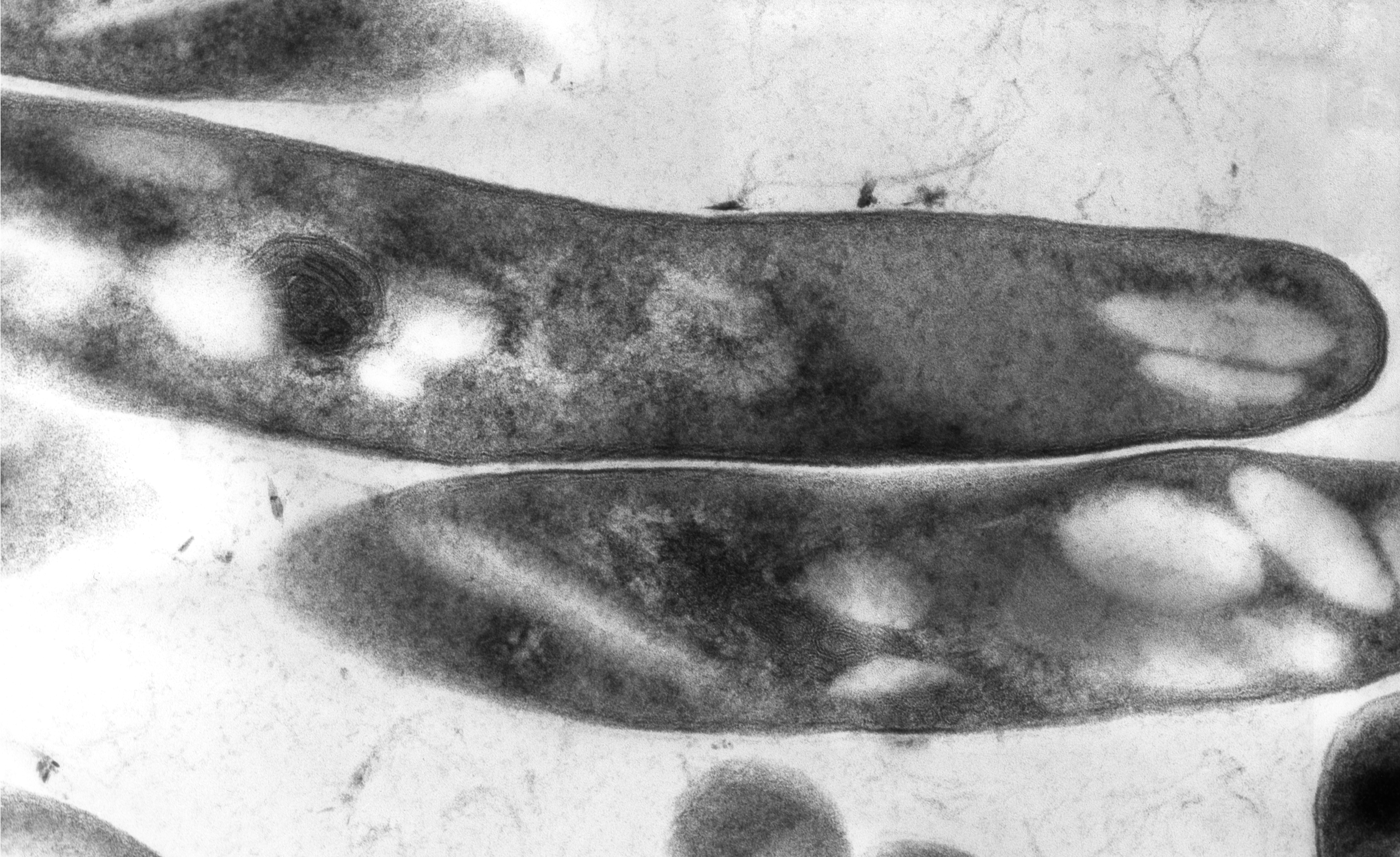
Mycobacterium is a genus of over 190 species in the phylum Actinomycetota, assigned its own family, Mycobacteriaceae. This genus includes pathogens known to cause serious diseases in mammals, including tuberculosis (M. tuberculosis) and leprosy (M. leprae) in humans. The Greek prefix myco- means 'fungus', alluding to this genus' mold-like colony surfaces.[3] Since this genus has cell walls with a waxy lipid-rich outer layer that contains high concentrations of mycolic acid,[4] acid-fast staining is used to emphasize their resistance to acids, compared to other cell types.[5]
Mycobacterial species are generally aerobic, non-motile, and capable of growing with minimal nutrients. The genus is divided based on each species' pigment production and growth rate.[6] While most Mycobacterium species are non-pathogenic, the genus' characteristic complex cell wall contributes to evasion from host defenses.[7]
Pathogenicity[edit]
Mycobacterium tuberculosis complex[edit]
Mycobacterium tuberculosis can remain latent in human hosts for decades after an initial infection, allowing it to continue infecting others. It has been estimated that a third of the world population has latent tuberculosis (TB).[22] M. tuberculosis has many virulence factors, which can be divided across lipid and fatty acid metabolism, cell envelope proteins, macrophage inhibitors, kinase proteins, proteases, metal-transporter proteins, and gene expression regulators.[23] Several lineages such as M. t. var. bovis (bovine TB) were considered separate species in the M, tuberculosis complex until they were finally merged into the main species in 2018.[24]
Leprosy[edit]
The development of Leprosy is caused by infection with either Mycobacterium leprae or Mycobacterium lepromatosis, two closely related bacteria. Roughly 200,000 new cases of infection are reported each year, and 80% of new cases are reported in Brazil, India, and Indonesia.[25] M. leprae infection localizes within the skin macrophages and Schwann cells found in peripheral nerve tissue.
The two most common methods for visualizing these acid-fast bacilli as bright red against a blue background are the Ziehl-Neelsen stain and modified Kinyoun stain. Fite's stain is used to color M. leprae cells as pink against a blue background. Rapid Modified Auramine O Fluorescent staining has specific binding to slowly-growing mycobacteria for yellow staining against a dark background. Newer methods include Gomori-Methenamine Silver staining and Perioidic Acid Schiff staining to color Mycobacterium avium complex (MAC) cells black and pink, respectively.[5]
While some mycobacteria can take up to eight weeks to grow visible colonies from a cultured sample, most clinically relevant species will grow within the first four weeks, allowing physicians to consider alternative causes if negative readings continue past the first month.[42] Growth media include Löwenstein–Jensen medium and mycobacteria growth indicator tube (MGIT).
Mycobacteriophages[edit]
Mycobacteria can be infected by mycobacteriophages, a class of viruses with high specificity for their targets. By hijacking the cellular machinery of mycobacteria to produce additional phages, such viruses can be used in phage therapy for eukaryotic hosts, as they would die alongside the mycobacteria. Since only some mycobacteriophages are capable of penetrating the M. tuberculosis membrane, the viral DNA may be delivered through artificial liposomes because bacteria uptake, transcribe, and translate foreign DNA into proteins.[43]
Mycosides[edit]
Mycosides are glycolipids isolated from Mycobacterium species with Mycoside A found in photochromogenic strains, Mycoside B in bovine strains, and Mycoside C in avian strains.[44] Different forms of Mycoside C have varying success as a receptor to inactivate mycobacteriophages.[45] Replacement of the gene encoding mycocerosic acid synthase in M. bovis prevents formation of mycosides.[46]

![Slant tubes of Löwenstein-Jensen medium.[note 1]](http://upload.wikimedia.org/wikipedia/commons/thumb/4/4a/Slant_tubes_of_L%C3%B6wenstein-Jensen_medium_with_control%2C_M_tuberculosis%2C_M_avium_and_M_gordonae.jpg/191px-Slant_tubes_of_L%C3%B6wenstein-Jensen_medium_with_control%2C_M_tuberculosis%2C_M_avium_and_M_gordonae.jpg)
