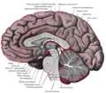
Pineal gland
The pineal gland (also known as the pineal body[1] or epiphysis cerebri) is a small endocrine gland in the brain of most vertebrates. In the darkness the pineal gland produces melatonin, a serotonin-derived hormone, which modulates sleep patterns following the diurnal cycles.[2] The shape of the gland resembles a pine cone, which gives it its name.[3] The pineal gland is located in the epithalamus, near the center of the brain, between the two hemispheres, tucked in a groove where the two halves of the thalamus join.[4][5] It is one of the neuroendocrine secretory circumventricular organs in which capillaries are mostly permeable to solutes in the blood.[6]
"Conarium" redirects here. For the video game, see Conarium (video game).Pineal gland
Neural ectoderm, roof of diencephalon
glandula pinealis
The pineal gland is present in almost all vertebrates, but is absent in protochordates in which there is a simple pineal homologue. The hagfish, considered as a primitive vertebrate, has a rudimentary structure regarded as the "pineal equivalent" in the dorsal diencephalon.[7] In some species of amphibians and reptiles, the gland is linked to a light-sensing organ, variously called the parietal eye, the pineal eye or the third eye.[8] Reconstruction of the biological evolution pattern suggests that the pineal gland was originally a kind of atrophied photoreceptor that developed into a neuroendocrine organ.
Ancient Greeks were the first to notice the pineal gland and believed it to be a valve, a guardian for the flow of pneuma. Galen in the 2nd century C.E. could not find any functional role and regarded the gland as a structural support for the brain tissue. He gave the name konario, meaning cone or pinecone, which during Renaissance was translated to Latin as pinealis. The 17th century philosopher René Descartes regarded the gland as having a mystical purpose, describing it as the "principal seat of the soul". In the mid-20th century, the real biological role as a neuroendocrine organ was established.[9]
Clinical significance[edit]
Calcification[edit]
Calcification of the pineal gland is typical in young adults, and has been observed in children as young as two years of age.[35] The internal secretions of the pineal gland are known to inhibit the development of the reproductive glands because when it is severely damaged in children, development of the sexual organs and the skeleton are accelerated.[36] Pineal gland calcification is detrimental to its ability to synthesize melatonin[37][38] and scientific literature presents inconclusive findings on whether it causes sleep problems.[39][40]
The calcified gland is often seen in skull X-rays.[35] Calcification rates vary widely by country and correlate with an increase in age, with calcification occurring in an estimated 40% of Americans by age seventeen.[35] Calcification of the pineal gland is associated with corpora arenacea, also known as "brain sand".
Tumors[edit]
Tumors of the pineal gland are called pinealomas. These tumors are rare and 50% to 70% are germinomas that arise from sequestered embryonic germ cells. Histologically they are similar to testicular seminomas and ovarian dysgerminomas.[41]
A pineal tumor can compress the superior colliculi and pretectal area of the dorsal midbrain, producing Parinaud's syndrome. Pineal tumors also can cause compression of the cerebral aqueduct, resulting in a noncommunicating hydrocephalus. Other manifestations are the consequence of their pressure effects and consist of visual disturbances, headache, mental deterioration, and sometimes dementia-like behaviour.[42]
These neoplasms are divided into three categories: pineoblastomas, pineocytomas, and mixed tumors, based on their level of differentiation, which, in turn, correlates with their neoplastic aggressiveness.[43] The clinical course of patients with pineocytomas is prolonged, averaging up to several years.[44] The position of these tumors makes them difficult to remove surgically.
Other animals[edit]
Nearly all vertebrate species possess a pineal gland. The most important exception is a primitive vertebrate, the hagfish. Even in the hagfish, however, there may be a "pineal equivalent" structure in the dorsal diencephalon.[7] A few more complex vertebrates have lost pineal glands over the course of their evolution.[46] The lamprey (another primitive vertebrate), however, does possess one.[47] The lancelet Branchiostoma lanceolatum, an early chordate which is a close relative to vertebrates, also lacks a recognizable pineal gland.[47] Protochordates in general do not have the distinct structure as an organ, but they have a mass of photoreceptor cells called lamellar body, which is regarded as a pineal homologue.[48][49]
The results of various scientific research in evolutionary biology, comparative neuroanatomy and neurophysiology have explained the evolutionary history (phylogeny) of the pineal gland in different vertebrate species. From the point of view of biological evolution, the pineal gland is a kind of atrophied photoreceptor. In the epithalamus of some species of amphibians and reptiles, it is linked to a light-sensing organ, known as the parietal eye, which is also called the pineal eye or third eye.[8] It is likely that the common ancestor of all vertebrates had a pair of photosensory organs on the top of its head, similar to the arrangement in modern lampreys.[50] In many lower vertebrates (such as species of fish, amphibians and lizards), the pineal gland is associated with parietal or pineal eye. In these animals, the parietal eye acts as a photoreceptor, and hence are also known as the third eye, and they can be seen on top of the head in some species.[51] Some extinct Devonian fishes have two parietal foramina in their skulls,[52][53] suggesting an ancestral bilaterality of parietal eyes. The parietal eye and the pineal gland of living tetrapods are probably the descendants of the left and right parts of this organ, respectively.[54]
During embryonic development, the parietal eye and the pineal organ of modern lizards[55] and tuataras[56] form together from a pocket formed in the brain ectoderm. The loss of parietal eyes in many living tetrapods is supported by developmental formation of a paired structure that subsequently fuses into a single pineal gland in developing embryos of turtles, snakes, birds, and mammals.[57]
The pineal organs of mammals fall into one of three categories based on shape. Rodents have more structurally complex pineal glands than other mammals.[58]
Crocodilians and some tropical lineages of mammals (some xenarthrans (sloths), pangolins, sirenians (manatees and dugongs), and some marsupials (sugar gliders)) have lost both their parietal eye and their pineal organ.[59][60][58] Polar mammals, such as walruses and some seals, possess unusually large pineal glands.[59]
All amphibians have a pineal organ, but some frogs and toads also have what is called a "frontal organ", which is essentially a parietal eye.[61]
Pinealocytes in many non-mammalian vertebrates have a strong resemblance to the photoreceptor cells of the eye. Evidence from morphology and developmental biology suggests that pineal cells possess a common evolutionary ancestor with retinal cells.[62]
Pineal cytostructure seems to have evolutionary similarities to the retinal cells of the lateral eyes.[62] Modern birds and reptiles express the phototransducing pigment melanopsin in the pineal gland. Avian pineal glands are thought to act like the suprachiasmatic nucleus in mammals.[63] The structure of the pineal eye in modern lizards and tuatara is analogous to the cornea, lens, and retina of the lateral eyes of vertebrates.[57]
In most vertebrates, exposure to light sets off a chain reaction of enzymatic events within the pineal gland that regulates circadian rhythms.[64] In humans and other mammals, the light signals necessary to set circadian rhythms are sent from the eye through the retinohypothalamic system to the suprachiasmatic nuclei (SCN) and the pineal gland.
The fossilized skulls of many extinct vertebrates have a pineal foramen (opening), which in some cases is larger than that of any living vertebrate.[65] Although fossils seldom preserve deep-brain soft anatomy, the brain of the Russian fossil bird Cerebavis cenomanica from Melovatka, about 90 million years old, shows a relatively large parietal eye and pineal gland.[66]
Society and culture[edit]
The notion of a "pineal-eye" is central to the philosophy of the French writer Georges Bataille, which is analyzed at length by literary scholar Denis Hollier in his study Against Architecture. In this work Hollier discusses how Bataille uses the concept of a "pineal-eye" as a reference to a blind-spot in Western rationality, and an organ of excess and delirium.[84] This conceptual device is explicit in his surrealist texts, The Jesuve and The Pineal Eye.[85]
In the late 19th century Madame Blavatsky, founder of theosophy, identified the pineal gland with the Hindu concept of the third eye, or the Ajna chakra. This association is still popular today.[9] The pineal gland has also featured in other religious contexts, such as in the Principia Discordia, which claims it can be used to contact the goddess of discord Eris.[86]
In the short story "From Beyond" by H. P. Lovecraft, a scientist creates an electronic device that emits a resonance wave, which stimulates an affected person's pineal gland, thereby allowing them to perceive planes of existence outside the scope of accepted reality, a translucent, alien environment that overlaps our own recognized reality. It was adapted as a film of the same name in 1986. The 2013 horror film Banshee Chapter is heavily influenced by this short story.
In Season 16, episode 6 of "American Dad" Steve tries to "astral project" using his pineal gland to help him understand the meaning of life. The episode is entitled "The Wondercabinet".
The pineal body is labeled in these images.


