Posterior cruciate ligament
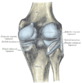
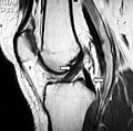
The posterior cruciate ligament (PCL) is a ligament in each knee of humans and various other animals. It works as a counterpart to the anterior cruciate ligament (ACL). It connects the posterior intercondylar area of the tibia to the medial condyle of the femur. This configuration allows the PCL to resist forces pushing the tibia posteriorly relative to the femur.
Posterior cruciate ligament
Antero-lateral aspect of medial femoral condyle
Posterolateral aspect of proximal tibia
ligamentum cruciatum posterius genus
The PCL and ACL are intracapsular ligaments because they lie deep within the knee joint. They are both isolated from the fluid-filled synovial cavity, with the synovial membrane wrapped around them. The PCL gets its name by attaching to the posterior portion of the tibia.[1]
The PCL, ACL, MCL, and LCL are the four main ligaments of the knee in primates.
Function[edit]
Although each PCL is a unified unit, they are described as separate anterolateral and posteromedial sections based on where each section's attachment site and function.[7] During knee joint movement, the PCL rotates [6][8] such that the anterolateral section stretches in knee flexion but not in knee extension and the posteromedial bundle stretches in extension rather than flexion.[4][9]
The function of the PCL is to prevent the femur from sliding off the anterior edge of the tibia and to prevent the tibia from displacing posterior to the femur. The posterior cruciate ligament is located within the knee. Ligaments are sturdy bands of tissues that connect bones. Similar to the anterior cruciate ligament, the PCL connects the femur to the tibia.
Other animals[edit]
In the quadruped stifle (analogous to the human knee), based on its anatomical position, it is referred to as the caudal cruciate ligament.[23]


