Skin condition
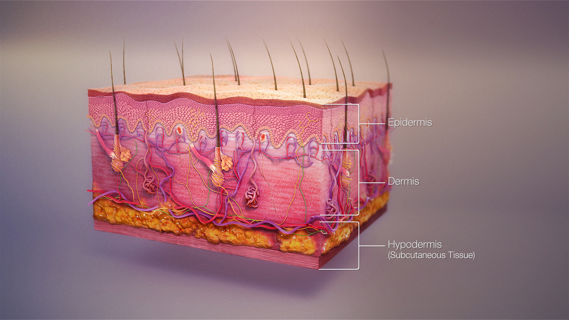
A skin condition, also known as cutaneous condition, is any medical condition that affects the integumentary system—the organ system that encloses the body and includes skin, nails, and related muscle and glands.[1] The major function of this system is as a barrier against the external environment.[2]
Skin condition
Cutaneous condition
Bacteria, viruses, fungi, parasites, insects, trauma, cancers, allergies, toxins, vitamin/nutritional deficiencies/excesses, prolonged pressure, impaired blood circulation, ingrown hairs or nails, autoimmune conditions, aging, sun exposure, radiation exposure, exposure to heat/cold, dryness, humidity, other organ damage or condition, substance usage or contact, hereditary conditions, etc.
Conditions of the human integumentary system constitute a broad spectrum of diseases, also known as dermatoses, as well as many nonpathologic states (like, in certain circumstances, melanonychia and racquet nails).[3][4] While only a small number of skin diseases account for most visits to the physician, thousands of skin conditions have been described.[5] Classification of these conditions often presents many nosological challenges, since underlying causes and pathogenetics are often not known.[6][7] Therefore, most current textbooks present a classification based on location (for example, conditions of the mucous membrane), morphology (chronic blistering conditions), cause (skin conditions resulting from physical factors), and so on.[8][9]
Clinically, the diagnosis of any particular skin condition begins by gathering pertinent information of the presenting skin lesion(s), including: location (e.g. arms, head, legs); symptoms (pruritus, pain); duration (acute or chronic); arrangement (solitary, generalized, annular, linear); morphology (macules, papules, vesicles); and color (red, yellow, etc.).[10] Some diagnoses may also require a skin biopsy which yields histologic information[11][12] that can be correlated with the clinical presentation and any laboratory data.[13][14] The introduction of cutaneous ultrasound has allowed the detection of cutaneous tumors, inflammatory processes, and skin diseases.[15]
Diagnoses[edit]
The physical examination of the skin and its appendages, as well as the mucous membranes, forms the cornerstone of an accurate diagnosis of cutaneous conditions.[29] Most of these conditions present with cutaneous surface changes termed "lesions," which have more or less distinct characteristics.[30] Often proper examination will lead the physician to obtain appropriate historical information and/or laboratory tests that are able to confirm the diagnosis.[29] Upon examination, the important clinical observations are the (1) morphology, (2) configuration, and (3) distribution of the lesion(s).[29] With regard to morphology, the initial lesion that characterizes a condition is known as the "primary lesion", and identification of such a lesions is the most important aspect of the cutaneous examination.[30] Over time, these primary lesions may continue to develop or be modified by regression or trauma, producing "secondary lesions".[1] However, with that being stated, the lack of standardization of basic dermatologic terminology has been one of the principal barriers to successful communication among physicians in describing cutaneous findings.[21] Nevertheless, there are some commonly accepted terms used to describe the macroscopic morphology, configuration, and distribution of skin lesions, which are listed below.[30]