Three prime untranslated region
In molecular genetics, the three prime untranslated region (3′-UTR) is the section of messenger RNA (mRNA) that immediately follows the translation termination codon. The 3′-UTR often contains regulatory regions that post-transcriptionally influence gene expression.
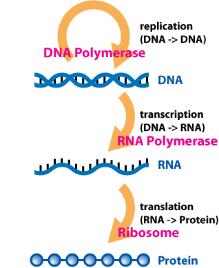
During gene expression, an mRNA molecule is transcribed from the DNA sequence and is later translated into a protein. Several regions of the mRNA molecule are not translated into a protein including the 5' cap, 5' untranslated region, 3′ untranslated region and poly(A) tail. Regulatory regions within the 3′-untranslated region can influence polyadenylation, translation efficiency, localization, and stability of the mRNA.[1][2] The 3′-UTR contains binding sites for both regulatory proteins and microRNAs (miRNAs). By binding to specific sites within the 3′-UTR, miRNAs can decrease gene expression of various mRNAs by either inhibiting translation or directly causing degradation of the transcript. The 3′-UTR also has silencer regions which bind to repressor proteins and will inhibit the expression of the mRNA.
Many 3′-UTRs also contain AU-rich elements (AREs). Proteins bind AREs to affect the stability or decay rate of transcripts in a localized manner or affect translation initiation. Furthermore, the 3′-UTR contains the sequence AAUAAA that directs addition of several hundred adenine residues called the poly(A) tail to the end of the mRNA transcript. Poly(A) binding protein (PABP) binds to this tail, contributing to regulation of mRNA translation, stability, and export. For example, poly(A) tail bound PABP interacts with proteins associated with the 5' end of the transcript, causing a circularization of the mRNA that promotes translation.
The 3′-UTR can also contain sequences that attract proteins to associate the mRNA with the cytoskeleton, transport it to or from the cell nucleus, or perform other types of localization. In addition to sequences within the 3′-UTR, the physical characteristics of the region, including its length and secondary structure, contribute to translation regulation. These diverse mechanisms of gene regulation ensure that the correct genes are expressed in the correct cells at the appropriate times.
Methods of study[edit]
Scientists use a number of methods to study the complex structures and functions of the 3′ UTR. Even if a given 3′-UTR in an mRNA is shown to be present in a tissue, the effects of localization, functional half-life, translational efficiency, and trans-acting elements must be determined to understand the 3′-UTR's full functionality.[7] Computational approaches, primarily by sequence analysis, have shown the existence of AREs in approximately 5 to 8% of human 3′-UTRs and the presence of one or more miRNA targets in as many as 60% or more of human 3′-UTRs. Software can rapidly compare millions of sequences at once to find similarities between various 3′ UTRs within the genome. Experimental approaches have been used to define sequences that associate with specific RNA-binding proteins; specifically, recent improvements in sequencing and cross-linking techniques have enabled fine mapping of protein binding sites within the transcript.[8] Induced site-specific mutations, for example those that affect the termination codon, polyadenylation signal, or secondary structure of the 3′-UTR, can show how mutated regions can cause translation deregulation and disease.[9] These types of transcript-wide methods should help our understanding of known cis elements and trans-regulatory factors within 3′-UTRs.
Future development[edit]
Despite current understanding of 3′-UTRs, they are still relative mysteries. Since mRNAs usually contain several overlapping control elements, it is often difficult to specify the identity and function of each 3′-UTR element, let alone the regulatory factors that may bind at these sites. Additionally, each 3′-UTR contains many alternative AU-rich elements and polyadenylation signals. These cis- and trans-acting elements, along with miRNAs, offer a virtually limitless range of control possibilities within a single mRNA.[7] Future research through the increased use of deep-sequencing based ribosome profiling will reveal more regulatory subtleties as well as new control elements and AUBPs.[1]