Katana VentraIP

Tibia

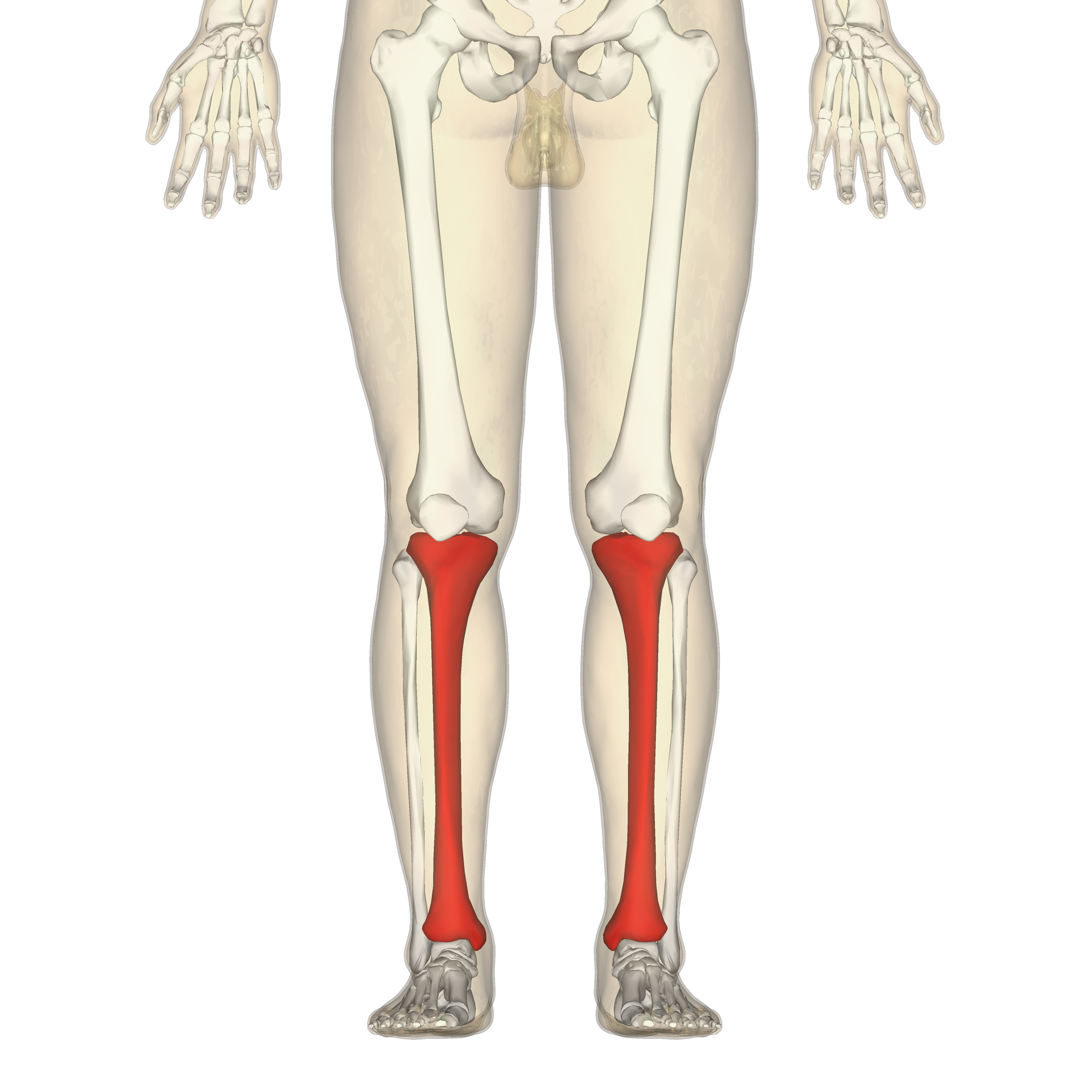
The tibia (/ˈtɪbiə/; pl.: tibiae /ˈtɪbii/ or tibias), also known as the shinbone or shankbone, is the larger, stronger, and anterior (frontal) of the two bones in the leg below the knee in vertebrates (the other being the fibula, behind and to the outside of the tibia); it connects the knee with the ankle. The tibia is found on the medial side of the leg next to the fibula and closer to the median plane. The tibia is connected to the fibula by the interosseous membrane of leg, forming a type of fibrous joint called a syndesmosis with very little movement. The tibia is named for the flute tibia. It is the second largest bone in the human body, after the femur. The leg bones are the strongest long bones as they support the rest of the body.

This article is about the human leg bone. For other uses, see Tibia (disambiguation).

The medial facet, oval in shape, is slightly concave from side to side, and from before backward.

The lateral, nearly circular, is concave from side to side, but slightly convex from before backward, especially at its posterior part, where it is prolonged on to the posterior surface for a short distance.

Clinical significance[edit]

Fracture[edit]

Fractures of the tibia can be divided into those that only involve the tibia; bumper fracture, Segond fracture, Gosselin fracture, toddler's fracture, and those including both the tibia and fibula; trimalleolar fracture, bimalleolar fracture, Pott's fracture.

Society and culture[edit]

In Judaism, the tibia, or shankbone, of a goat or sheep is used in the Passover Seder plate.

Other animals[edit]

The structure of the tibia in most other tetrapods is essentially similar to that in humans. The tuberosity of the tibia, a crest to which the patellar ligament attaches in mammals, is instead the point for the tendon of the quadriceps muscle in reptiles, birds, and amphibians, which have no patella.[9]

Shape of right tibia

Shape of right tibia

3D image

3D image

Longitudinal section of tibia showing interior

Longitudinal section of tibia showing interior

Right knee-joint. Anterior view.

Right knee-joint. Anterior view.

Right knee joint from the front, showing interior ligaments

Right knee joint from the front, showing interior ligaments

Left knee joint from behind, showing interior ligaments

Left knee joint from behind, showing interior ligaments

Left talocrural joint

Left talocrural joint

Coronal section through right talocrural and talocalcaneal joints

Coronal section through right talocrural and talocalcaneal joints

Dorsum of Foot. Ankle joint. Deep dissection

Dorsum of Foot. Ankle joint. Deep dissection

Dorsum of Foot. Ankle joint. Deep dissection

Dorsum of Foot. Ankle joint. Deep dissection

Ankle joint. Deep dissection. Anterior view

Ankle joint. Deep dissection. Anterior view

Bones of the right leg. Anterior surface

Bones of the right leg. Anterior surface

Bones of the right leg. Posterior surface

Bones of the right leg. Posterior surface

Dorsum of Foot. Ankle joint. Deep dissection.

Dorsum of Foot. Ankle joint. Deep dissection.

Ankle joint. Deep dissection.

Ankle joint. Deep dissection.

Ankle joint. Deep dissection.

Ankle joint. Deep dissection.

Ankle joint. Deep dissection.

Ankle joint. Deep dissection.

Ankle joint. Deep dissection.

Ankle joint. Deep dissection.

Tibia Anatomy

Shin splints

Squatting facets