Visual cortex
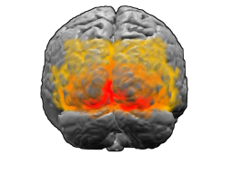
The visual cortex of the brain is the area of the cerebral cortex that processes visual information. It is located in the occipital lobe. Sensory input originating from the eyes travels through the lateral geniculate nucleus in the thalamus and then reaches the visual cortex. The area of the visual cortex that receives the sensory input from the lateral geniculate nucleus is the primary visual cortex, also known as visual area 1 (V1), Brodmann area 17, or the striate cortex. The extrastriate areas consist of visual areas 2, 3, 4, and 5 (also known as V2, V3, V4, and V5, or Brodmann area 18 and all Brodmann area 19).[1]
Visual cortex
Both hemispheres of the brain include a visual cortex; the visual cortex in the left hemisphere receives signals from the right visual field, and the visual cortex in the right hemisphere receives signals from the left visual field.
Introduction[edit]
The primary visual cortex (V1) is located in and around the calcarine fissure in the occipital lobe. Each hemisphere's V1 receives information directly from its ipsilateral lateral geniculate nucleus that receives signals from the contralateral visual hemifield.
Neurons in the visual cortex fire action potentials when visual stimuli appear within their receptive field. By definition, the receptive field is the region within the entire visual field that elicits an action potential. But, for any given neuron, it may respond best to a subset of stimuli within its receptive field. This property is called neuronal tuning. In the earlier visual areas, neurons have simpler tuning. For example, a neuron in V1 may fire to any vertical stimulus in its receptive field. In the higher visual areas, neurons have complex tuning. For example, in the inferior temporal cortex (IT), a neuron may fire only when a certain face appears in its receptive field.
Furthermore, the arrangement of receptive fields in V1 is retinotopic, meaning neighboring cells in V1 have receptive fields that correspond to adjacent portions of the visual field. This spatial organization allows for a systematic representation of the visual world within V1. Additionally, recent studies have delved into the role of contextual modulation in V1, where the perception of a stimulus is influenced not only by the stimulus itself but also by the surrounding context, highlighting the intricate processing capabilities of V1 in shaping our visual experiences.[2]
The visual cortex receives its blood supply primarily from the calcarine branch of the posterior cerebral artery.
The size of V1, V2, and V3 can vary three-fold, a difference that is partially inherited.[3]