Abdominal aorta
In human anatomy, the abdominal aorta is the largest artery in the abdominal cavity. As part of the aorta, it is a direct continuation of the descending aorta (of the thorax).[1]
This article uses anatomical terminology.Abdominal aorta
aorta abdominalis,
pars abdominalis aortae
Contrast enhanced MRA of the abdominal aorta demonstrating normal paired arteries.

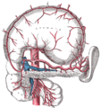
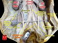
The celiac artery and its branches; the stomach has been raised and the peritoneum removed.
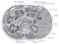
Transverse section through the middle of the first lumbar vertebra, showing the relations of the pancreas.
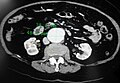
CT scan showing the liver and a kidney

A transverse contrast enhanced CT scan demonstrating an abdominal aortic aneurysm of 4.8 by 3.8 cm
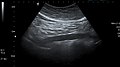
The standard aortic measurement on abdominal ultrasonography, such as used for abdominal aortic aneurysms, is between the outer margins of the aortic wall.[3]
![The standard aortic measurement on abdominal ultrasonography, such as used for abdominal aortic aneurysms, is between the outer margins of the aortic wall.[3]](http://upload.wikimedia.org/wikipedia/commons/thumb/7/7b/Ultrasonographic_measurement_of_aortic_diameter_at_the_navel.svg/120px-Ultrasonographic_measurement_of_aortic_diameter_at_the_navel.svg.png)
Abdominal aorta
Abdominal aorta ultrasound