Mammography
Mammography (also called mastography: DICOM modality = MG) is the process of using low-energy X-rays (usually around 30 kVp) to examine the human breast for diagnosis and screening. The goal of mammography is the early detection of breast cancer, typically through detection of characteristic masses or microcalcifications.
See also: Breast cancer screening
As with all X-rays, mammograms use doses of ionizing radiation to create images. These images are then analyzed for abnormal findings. It is usual to employ lower-energy X-rays, typically Mo (K-shell X-ray energies of 17.5 and 19.6 keV) and Rh (20.2 and 22.7 keV) than those used for radiography of bones. Mammography may be 2D or 3D (tomosynthesis), depending on the available equipment and/or purpose of the examination. Ultrasound, ductography, positron emission mammography (PEM), and magnetic resonance imaging (MRI) are adjuncts to mammography. Ultrasound is typically used for further evaluation of masses found on mammography or palpable masses that may or may not be seen on mammograms. Ductograms are still used in some institutions for evaluation of bloody nipple discharge when the mammogram is non-diagnostic. MRI can be useful for the screening of high-risk patients, for further evaluation of questionable findings or symptoms, as well as for pre-surgical evaluation of patients with known breast cancer, in order to detect additional lesions that might change the surgical approach (for example, from breast-conserving lumpectomy to mastectomy).
In 2023, the U.S. Preventive Services Task Force issued a draft recommendation statement that all women should receive a screening mammography every two years from age 40 to 74.[1][2]The American College of Radiology and American Cancer Society recommend yearly screening mammography starting at age 40.[3] The Canadian Task Force on Preventive Health Care (2012) and the European Cancer Observatory (2011) recommend mammography every 2 to 3 years between ages 50 and 69.[4][5] These task force reports point out that in addition to unnecessary surgery and anxiety, the risks of more frequent mammograms include a small but significant increase in breast cancer induced by radiation.[6][7] Additionally, mammograms should not be performed with increased frequency in patients undergoing breast surgery, including breast enlargement, mastopexy, and breast reduction.[8]
Types[edit]
Digital[edit]
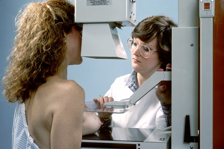
Digital mammography is a specialized form of mammography that uses digital receptors and computers instead of X-ray film to help examine breast tissue for breast cancer.[9] The electrical signals can be read on computer screens, permitting more manipulation of images to allow radiologists to view the results more clearly .[9][10] Digital mammography may be "spot view", for breast biopsy,[11] or "full field" (FFDM) for screening.[9]
Digital mammography is also utilized in stereotactic biopsy. Breast biopsy may also be performed using a different modality, such as ultrasound or magnetic resonance imaging (MRI).
While radiologists[12] had hoped for more marked improvement, the effectiveness of digital mammography was found comparable to traditional X-ray methods in 2004, though there may be reduced radiation with the technique and it may lead to fewer retests.[9] Specifically, it performs no better than film for post-menopausal women, who represent more than three-quarters of women with breast cancer.[13] The U.S. Preventive Services Task Force concluded that there was insufficient evidence to recommend for or against digital mammography.[14]
Digital mammography is a NASA spin-off, utilizing technology developed for the Hubble Space Telescope.[15] As of 2007, about 8% of American screening centers used digital mammography. Around the globe, systems by Fujifilm Corporation are the most widely used. In the United States, GE's digital imaging units typically cost US$300,000 to $500,000, far more than film-based imaging systems.[13] Costs may decline as GE begins to compete with the less expensive Fuji systems.[13]
3D mammography[edit]
Three-dimensional mammography, also known as digital breast tomosynthesis (DBT), tomosynthesis, and 3D breast imaging, is a mammogram technology that creates a 3D image of the breast using X-rays. When used in addition to usual mammography, it results in more positive tests.[16] Cost effectiveness is unclear as of 2016.[17] Another concern is that it more than doubles the radiation exposure.[18]
When to start screening[edit]
In 2014, the Surveillance, Epidemiology, and End Results Program of the National Institutes of Health reported the occurrence rates of breast cancer based on 1000 women in different age groups.[28] In the 40–44 age group, the incidence was 1.5 and in the 45–49 age group, the incidence was 2.3.[28] In the older age groups, the incidence was 2.7 in the 50–54 age group and 3.2 in the 55–59 age group.[28]
While screening between ages 40 and 50 is somewhat controversial, the preponderance of the evidence indicates that there is a benefit in terms of early detection. Currently, the American Cancer Society, the American Congress of Obstetricians and Gynecologists (ACOG), the American College of Radiology, and the Society of Breast Imaging encourage annual mammograms beginning at age 40.[29][30][31]
The National Cancer Institute encourages mammograms every one to two years for women ages 40 to 49.[32] In 2023, United States Preventive Services Task Force (USPSTF) revised the recommendation that women and transgender men undergo biennial mammograms starting at the age of 40, rather than the previously suggested age of 50.[33] This adjustment is prompted by the increasing incidence of breast cancer in the 40 to 49 age group over the past decade.
In contrast, the American College of Physicians, a large internal medicine group, has recently encouraged individualized screening plans as opposed to wholesale biannual screening of women aged 40 to 49.[34] The American Cancer Society recommendations for women at average risk for breast cancer is a yearly mammogram from age 45 to 54 with an optional yearly mammogram from age 40 to 44.[35]
Women who are at high risk for early-onset breast cancer have separate recommendations for screening. These include those who:
The American College of Radiology recommends these individuals to get annual mammography starting at the age of 30. Those with a history of chest radiation therapy before age 30 should start annually at age 25 of 8 years after their latest therapy (whichever is latest).[37] The American Cancer Society also recommends women at high risk should get a mammogram and breast MRI every year beginning at age 30 or an age recommended by their healthcare provider.[35]
The National Comprehensive Cancer Network (NCCN) advocates screening for women who possess a BRCA1 or BRCA2 mutation or have a first-degree relative with such a mutation, even in the absence of the patient being tested for BRCA1/2 mutations. For women at high risk, NCCN recommends undergoing an annual mammogram and breast MRI between the ages of 25 and 40, considering the specific gene mutation type and/or the youngest age of breast cancer occurrence in the family. Additionally, NCCN suggests that high-risk women undergo clinical breast exams every 6 to 12 months starting at age 25. These individuals should also engage in discussions with healthcare providers to assess the advantages and disadvantages of 3D mammography and acquire knowledge on detecting changes in their breasts.[38]
Adverse effects[edit]
Radiation[edit]
The radiation exposure associated with mammography is a potential risk of screening, which appears to be greater in younger women. In scans where women receive 0.25–20 Gray (Gy) of radiation, they have more of an elevated risk of developing breast cancer.[39] A study of radiation risk from mammography concluded that for women 40 years of age and older, the risk of radiation-induced breast cancer was minuscule, particularly compared with the potential benefit of mammographic screening, with a benefit-to-risk ratio of 48.5 lives saved for each life lost due to radiation exposure.[40] This also correlates to a decrease in breast cancer mortality rates by 24%.[39]
Pain[edit]
The mammography procedure can be painful. Reported pain rates range from 6–76%, with 23–95% experiencing pain or discomfort.[41] Experiencing pain is a significant predictor in women not re-attending screening.[42] There are few proven interventions to reduce pain in mammography, but evidence suggests that giving women information about the mammography procedure prior to it taking place may reduce the pain and discomfort experienced.[43] Furthermore, research has found that standardised compression levels can help to reduce patients' pain while still allowing for optimal diagnostic images to be produced.[44]
History[edit]
As a medical procedure that induces ionizing radiation, the origin of mammography can be traced to the discovery of X-rays by Wilhelm Röntgen in 1895.
In 1913, German surgeon Albert Salomon performed a mammography study on 3,000 mastectomies, comparing X-rays of the breasts to the actual removed tissue, observing specifically microcalcifications.[57][58] By doing so, he was able to establish the difference as seen on an X-ray image between cancerous and non-cancerous tumors in the breast.[58] Salomon's mammographs provided substantial information about the spread of tumors and their borders.[59]
In 1930, American physician and radiologist Stafford L. Warren published "A Roentgenologic Study of the Breast",[60] a study where he produced stereoscopic X-rays images to track changes in breast tissue as a result of pregnancy and mastitis.[61][62] In 119 women who subsequently underwent surgery, he correctly found breast cancer in 54 out of 58 cases.[61]
As early as 1937, Jacob Gershon-Cohen developed a form a mammography for a diagnostic of breast cancer at earlier stages to improve survival rates.[63] In 1949, Raul Leborgne sparked renewed enthusiasm for mammography by emphasizing the importance of technical proficiency in patient positioning and the adoption of specific radiological parameters. He played a pioneering role in elevating imaging quality while placing particular emphasis on distinguishing between benign and malignant calcifications.[64] In the early 1950s, Uruguayan radiologist Raul Leborgne developed the breast compression technique to produce better quality images, and described the differences between benign and malign microcalcifications.[65]
In 1956, Gershon-Cohen conducted clinical trails on over 1,000 asymptomatic women at the Albert Einstein Medical Center on his screening technique,[63] and the same year, Robert Egan at the University of Texas M.D. Anderson Cancer Center combined a technique of low kVp with high mA and single emulsion films developed by Kodak to devise a method of screening mammography. He published these results in 1959 in a paper, subsequently vulgarized in a 1964 book called Mammography.[66] The "Egan technique", as it became known, enabled physicians to detect calcification in breast tissue;[67] of the 245 breast cancers that were confirmed by biopsy among 1,000 patients, Egan and his colleagues at M.D. Anderson were able to identify 238 cases by using his method, 19 of which were in patients whose physical examinations had revealed no breast pathology.
Use of mammography as a screening technique spread clinically after a 1966 study demonstrating the impact of mammograms on mortality and treatment led by Philip Strax. This study, based in New York, was the first large-scale randomized controlled trial of mammography screening.[68][69]
In 1985, László Tabár and colleagues documented findings from mammographic screening involving 134,867 women aged 40 to 79. Using a single mediolateral oblique image, they reported a 31% reduction in mortality.[64] Dr. Tabár has since written many publications promoting mammography in the areas of epidemiology, screening, early diagnosis, and clinical-radiological-pathological correlation.
Society and culture[edit]
Attendance[edit]
Many factors affect how many people attend breast cancer screenings. For example, people from minority ethnic communities are also less likely to attend cancer screening. In the UK, women of South Asian heritage are the least likely to attend breast cancer screening. Research is still needed to identify specific barriers for the different South Asian communities. For example, a study showed that British-Pakistani women faced cultural and language barriers and were not aware that breast screening takes place in a female-only environment.[98][99][100]
People with mental illnesses are also less likely to attend cancer screening appointments.[101][102] In Northern Ireland women with mental health problems were shown to be less likely to attend screening for breast cancer, than women without. The lower attendance numbers remained the same even when marital status and social deprivation were taken into account.[103][104]
Regulation[edit]
Mammography facilities in the United States and its territories (including military bases) are subject to the Mammography Quality Standards Act (MQSA). The act requires annual inspections and accreditation every three years through an FDA-approved body. Facilities found deficient during the inspection or accreditation process can be barred from performing mammograms until corrective action has been verified or, in extreme cases, can be required to notify past patients that their exams were sub-standard and should not be relied upon.[105]
At this time, MQSA applies only to traditional mammography and not to related scans, such as breast ultrasound, stereotactic breast biopsy, or breast MRI.
As of September 10, 2024, the MQSA requires that all patients be notified of their breast density ("dense" or "not dense") in their mammogram reports.[106][107]
Research[edit]
Artificial intelligence (AI) algorithms[edit]
Recently, artificial intelligence (AI) programs have been developed to utilize features from screening mammography images to predict breast cancer risk. A systematic review of 16 retrospective study designs comparing median maximum AUC found that artificial intelligence had a comparable or better accuracy (AUC = 0.72) of predicting breast cancer than clinical risk factors alone (AUC = 0.61), suggesting a transition from clinical risk factor-based to AI image-based risk models may lead to more accurate and personalized risk-based screening approaches.[108]
Another study of 32 published papers involving 23,804 mammograms and various machine learning methods (CNN, ANN, and SVM) found promising results in the ability to assist clinicians in large-scale population-based breast cancer screening programs.[109]
Alternative examination methods[edit]
For patients who do not want to undergo mammography, MRI and also breast computed tomography (also called breast CT) offer a painless alternative. Whether the respective method is suitable depends on the clinical picture; it is decided by the physician.