Aortic arch
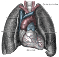
The aortic arch, arch of the aorta, or transverse aortic arch (English: /eɪˈɔːrtɪk/[1][2]) is the part of the aorta between the ascending and descending aorta. The arch travels backward, so that it ultimately runs to the left of the trachea.
For the embryological structure, see Aortic arches.Aortic arch
arcus aortae
Clinical significance[edit]
The aortic knob is the prominent shadow of the aortic arch on a frontal chest radiograph.[18]
Aortopexy is a surgical procedure in which the aortic arch is fixed to the sternum in order to keep the trachea open.
Aortic isthmus is the relatively fixed part of the aortic arch. It is prone to shearing force and trauma that can cause it to tear and result in massive bleeding.[3]

