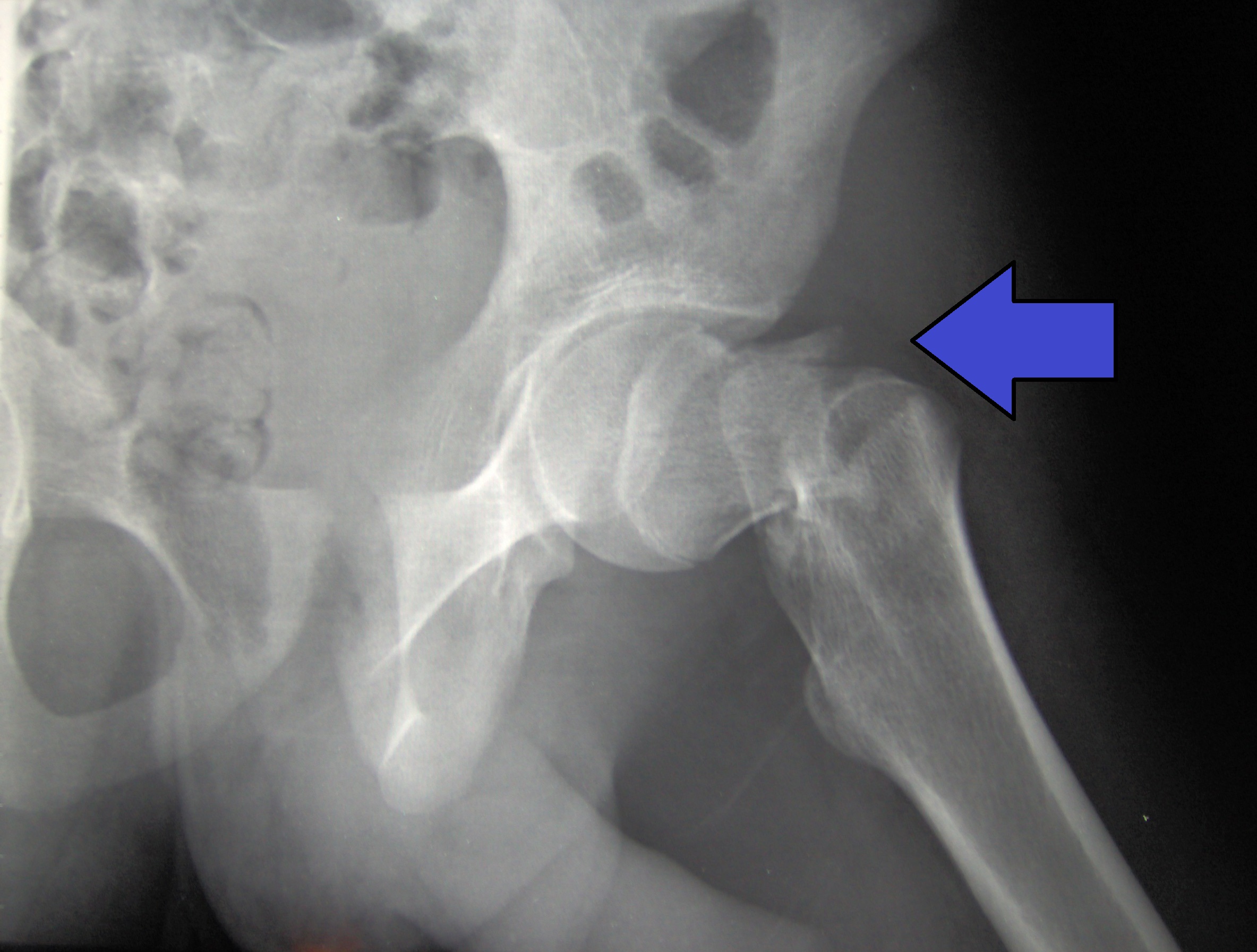
Hip fracture
A hip fracture is a break that occurs in the upper part of the femur (thigh bone), at the femoral neck or (rarely) the femoral head.[2] Symptoms may include pain around the hip, particularly with movement, and shortening of the leg.[2] Usually the person cannot walk.[3]
This article is about fractures of the proximal femur. For fractures of the nonacetabular portion of the hip bone, see Pelvic fracture. For fractures of the acetabulum, see Acetabular fracture.Hip fracture
Proximal femur fracture;[1] femoral neck fracture [main hyponym]; femoral head fracture [other hyponym]
Intracapsular, extracapsular (intertrochanteric, subtrochanteric, greater trochanteric, lesser trochanteric)[1]
Osteoporosis, taking many medications, alcohol use, metastatic cancer[2][1]
Improved lighting, removal of loose rugs, exercise, treatment of osteoporosis[1]
Surgery[1]
~15% of women at some point[1]
A hip fracture is usually a femoral neck fracture. Such fractures most often occur as a result of a fall.[3] (Femoral head fractures are a rare kind of hip fracture that may also be the result of a fall but are more commonly caused by more violent incidents such as traffic accidents.) Risk factors include osteoporosis, taking many medications, alcohol use, and metastatic cancer.[2][1] Diagnosis is generally by X-rays.[2] Magnetic resonance imaging, a CT scan, or a bone scan may occasionally be required to make the diagnosis.[3][2]
Pain management may involve opioids or a nerve block.[1][4] If the person's health allows, surgery is generally recommended within two days.[2][1] Options for surgery may include a total hip replacement or stabilizing the fracture with screws.[2] Treatment to prevent blood clots following surgery is recommended.[1]
About 15% of women break their hip at some point in life;[1] women are more often affected than men.[1] Hip fractures become more common with age.[1] The risk of death in the year following a fracture is about 20% in older people.[1][3]
Hip fracture following a fall is likely to be a pathological fracture. The most common causes of weakness in bone are:
Diagnosis[edit]
Imaging[edit]
Typically, radiographs are taken of the hip from the front (AP view), and side (lateral view). Frog leg views are to be avoided, as they may cause severe pain and further displace the fracture.[5] In situations where a hip fracture is suspected but not obvious on x-ray, an MRI is the next test of choice. If an MRI is not available or the patient can not be placed into the scanner a CT may be used as a substitute. MRI sensitivity for radiographically occult fracture is greater than CT. Bone scan is another useful alternative however substantial drawbacks include decreased sensitivity, early false negative results and decreased conspicuity of findings due to age-related metabolic changes in the elderly.[16]
A case demonstrating a possible order of imaging in initially subtle findings:
Prevention[edit]
The majority of hip fractures are the result of a fall, particularly in the elderly. Therefore, identifying why the fall occurred, and implementing treatments or changes, is key to reducing the occurrence of hip fractures. Multiple contributing factors are often identified.[24] These can include environmental factors and medical factors (such as postural hypotension or co-existing disabilities from disease such as Stroke or Parkinson's disease which cause visual and/or balance impairments). A recent study has identified a high incidence of undiagnosed cervical spondylotic myelopathy (CSM) amongst patients with a hip fracture.[25] This is relatively unrecognised consequent of CSM.[26]
Additionally, there is some evidence to systems designed to offer protection in the case of a fall. Hip protectors, for example appear to decrease the number of hip fractures among the elderly, but they are often not used.[27]